A research team led by engineers at the University of Leeds has achieved a breakthrough by producing high-resolution 3D ultrasound images using a probe inserted deep into the gastrointestinal tract for the first time.
This advancement could revolutionize cancer diagnosis and treatment through the use of “virtual biopsies”—non-invasive imaging techniques that deliver immediate diagnostic information. These scans could allow doctors to detect, assess, and even begin treating lesions in a single procedure, removing the need for traditional biopsies.
The unexpected secret to this achievement was the adoption of a little-known 3D geometric form called the oloid. This shape enabled the magnetic medical robot to gain a rolling movement, previously unattainable, which is crucial for precise internal navigation and imaging.
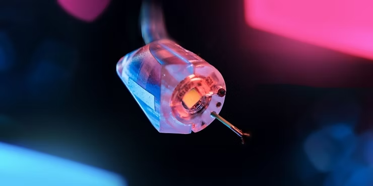
A study published on March 26 in Science Robotics details how the team incorporated the oloid’s unique rolling capability into a newly developed magnetic flexible endoscope (MFE), which was also equipped with a compact, high-frequency ultrasound device to generate detailed 3D visuals of internal tissues.
This innovative technology is the result of a collaborative effort among engineers, scientists, and medical professionals from the University of Leeds, the University of Glasgow, and the University of Edinburgh. The team at Leeds spearheaded the robotics development and integration of the imaging probe, while Glasgow and Edinburgh focused on providing the ultrasound device and leading the imaging research.
Professor Pietro Valdastri, Chair in Robotics and Autonomous Systems and Director of the STORM Lab at Leeds, led the study. He stated: “This is the first time we’ve been able to generate a 3D ultrasound image from a probe positioned deep within the gastrointestinal tract – a breakthrough that has never been achieved before.
“This method allows real-time tissue analysis and diagnosis of colorectal cancer right where it’s found. Currently, diagnosing this type of cancer involves removing a tissue sample and sending it to a lab, with results taking up to three weeks.”
The imaging system—a 28 MHz micro-ultrasound array—captures high-resolution 3D visuals of the scanned area. From these virtual reconstructions, doctors can produce cross-sectional images that resemble those created through traditional biopsies, where tissue samples are sliced and examined under a microscope.
High-frequency, or high-resolution, ultrasound differs from the standard ultrasound typically used to examine a fetus or internal organs. The probe used in this study offers much finer detail, allowing visualization at a microscopic scale, including individual tissue layers.
Although 3D ultrasound imaging is already possible within blood vessels and the rectum, this research expands the potential for performing detailed 3D scans further inside the gastrointestinal tract.
Postgraduate researcher Nikita Greenidge, the lead author of the study and a member of Leeds’ STORM Lab in the School of Electronic and Electrical Engineering, explained: “By merging our cutting-edge robotics with ultrasound imaging technology, we’ve advanced beyond conventional colonoscopy.
This development allows physicians to both diagnose and treat in a single session—eliminating delays between identifying a problem and taking action. This makes the experience more comfortable for patients, shortens waiting times, reduces the need for follow-up procedures, and eases the stress of waiting for potential cancer diagnoses.”
She further noted: “Colorectal cancer remains one of the leading causes of cancer deaths both in the UK and globally, yet early detection greatly improves treatment outcomes. Our approach offers a minimally invasive method that could enhance early diagnosis and, potentially, enable precise ultrasound-activated drug delivery in the future for more effective therapies.”
The study was supported by funding from UKRI’s Engineering and Physical Sciences Research Council (EPSRC), the European Commission (EC), the European Research Council (ERC), and the NIHR Leeds Biomedical Research Centre. It revealed that using the oloid—a unique shape formed by two perpendicular intersecting circles—significantly improved the flexibility, imaging capabilities, and autonomy of magnetic flexible endoscopes and magnetic medical robots in general.
The oloid magnetic endoscope (OME) was created using 3D-printed resin and measures just 21 mm in diameter—about the size of a one-pence coin—making it small enough to roll effectively while remaining suitable for clinical procedures like colonoscopies. Its mobility was evaluated on various surfaces designed to mimic the internal structures of the colon, esophagus, and stomach.
To move the technology closer to human trials, researchers began with tests in an artificial colon and then progressed to studies in pigs—an essential step in meeting regulatory standards for medical device approval. They used a robotically controlled external permanent magnet system, previously developed at Leeds, to steer the OME. This system allowed for both joystick-based and autonomous operation, guided by visuals from an onboard camera and a magnetic tracking system.
The study demonstrated that the system could:
- Effectively carry out controlled rolling and sweeping motions within the colon.
- Produce high-resolution 3D ultrasound images for precise diagnosis.
- Detect lesions in gastrointestinal tissues, highlighting its promise for advanced imaging and early disease identification.
Ms. Greenidge explained that although this research focused on the colon, the unique rolling capabilities of the oloid shape could be utilized in a range of magnetic medical robots, potentially broadening its use to other parts of the body.

The science of magnetic robots
Professor Sandy Cochran of the University of Glasgow’s Centre for Medical and Industrial Ultrasonics, who led the ultrasound portion of the project, noted that ultrasound is a safe, cost-effective tool that can be deployed exactly where it’s needed. By marrying medical ultrasound with state‑of‑the‑art robotics, she said the team aims to drive groundbreaking improvements in cancer detection, treatment, and overall patient care.
The researchers believe these innovations could fundamentally reshape endoscopy: clinicians would be freed to focus on critical diagnostic and therapeutic decisions, while autonomous systems handle routine navigation and operational tasks.
They also suggest that the OME’s superior maneuverability and imaging precision might help close gender gaps in colonoscopy outcomes—standard flexible scopes often prove more difficult to use in women, leading to higher rates of incomplete exams.
Jane Nicholson, Executive Director of Research at EPSRC, added that advances in cutting‑edge technologies are enabling the creation of rapid, non‑invasive solutions with the potential to revolutionize cancer diagnosis and treatment.
By enhancing the accuracy and control of procedures for common cancers like colorectal cancer, this multidisciplinary team’s work could usher in major breakthroughs in cancer detection and treatment.